
Our goal is to measure the forces and work required to move vesicles in live cells and to compare these to the limits established for kinesin in solution. Of particular interest is fast transport of vesicles toward the + ends of microtubules in axons by conventional kinesin. We are investigating whether kinesin in PC12 neurites or the axons of chicken neurons works the same as it does in buffer, where it has been extensively characterized. One expects highly extended cells, such as neurons, to be particularly sensitive to the energy costs of vesicle transport.
When the tail of a single kinesin molecule is attached to a glass or latex bead, it can pull the bead along a microtubule with a maximum force of 6.5 pNwithout stalling, as measured with an optical trap. One ATP is hydrolyzed per 8 nm step, and each step takes 50 microseconds. About 100 such steps occur per second during processive movement. In a cell, the vesicle is in cytoplasm, so the load on the motor is viscoelastic drag. The viscous part of this load differs by a factor of 10,000-100,000 from the viscous load in an optical trap, since the viscosity of water is .001 Pa.s and the zero-frequency viscosity of the cytoplasm of a "typical" cell, such as COS7, is roughly 50 Pa.s. Does kinesin develop the same force in the cell as it does in buffer?
We have computed the force and work that kinesin must exert in COS7 cells, assuming that kinesin takes the same 50-microsecond, 8-nm steps in the cell as it does in buffer. The required viscoelastic properties of cytoplasm were taken from measurements reported by Yamada, Wirtz, and Kuo for COS7 cells. Using these data and the theory of Thomas, Walters, Hwang, Litt, and Forsman for the forces on a sphere in a viscoelastic fluid, where Stokes' Law is invalid, we showed that the force required to move a 200-nm bead exceeded 75 pN at all times and peaked to 92 pN during the step, well beyond the measured capabilities of a single kinesin molecule. The computed work per 8-nm step in COS7 cytoplasm was 7 +/- 5 ATP, whereas in solution 1 ATP is consumed per 8-nm step. For details, see Biophysical Journal Vol 82, p.1784-1790 (2002).
We are now carrying out experiments to characterize vesicle transport and to measure the forces required to transport vesicles in PC12 cells. Instead of directly measuring the forces on 200-1000 nm endogenous vesicles, we are placing 500 nm superparamagnetic beads into the cells to mimic the vesicles. We have constructed a small magnet which can apply the required fields and field gradients to cells in a DIC microscope. Well-defined forces can be applied to a bead without damage to the cell containing the bead.
Shown below are DIC and fluorescence images of a magnetic bead in a PC12 cell. The bead was coated with fibronectin to enhance uptake. Mouse-over to see the same cell in flourescence.
Figure 1: PC12 neurites after 5 days of growth with NGF, recorded in DIC and fluorescence (on mouse rollover). Zeiss Axioplan, 40X objective, 2X Optovar, Hamamatsu Orca 12 bit gray-scale camera. Software: Image Pro Plus 4.0.
To know how fast to move the magnetic beads in PC12 cells (Fig. 1 above), the velocity of endogenous vesicles must be determined. Shown below are movies of differentiated PC12 cells and vesicles within them. Images obtained by DIC microscopy using a Nikon E600FN at 60X/W objective, Hamamatsu Orca 12-bit cooled CCD camera, and Matrox Genesis image-processing board. Vesicles are made more visible (click on inset) by subtracting a background image from the raw DIC image.
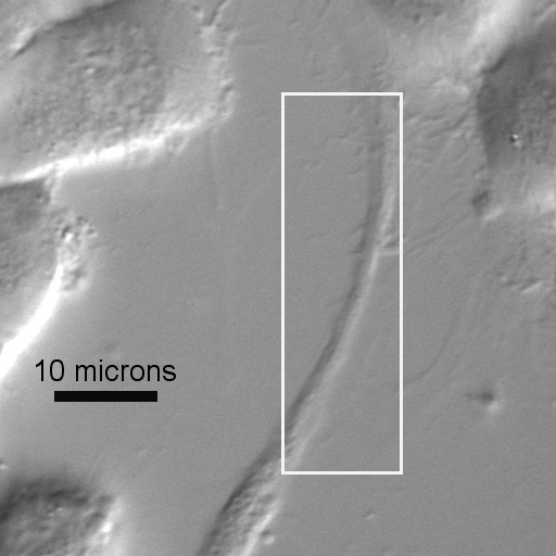
Fig. 2. Wide-field image of live PC12 cells and movie of intracellular vesicles travelling within one neurite. Click on the AOI to see a movie of vesicles being transported along the neurite. Wide-field image acquired by DIC microscopy using Nikon E600FN, 60X/W, Hamamatsu Orca camera. Movie shows background-subtracted DIC images.
![]()
Fig. 3. Wide-field image of live PC12 cells and background-subtracted frames at t=0, 1, and 3 s. The arrow marks a moving vesicle. Click on the AOI to in the wide-field image to see a vesicle transport more clearly. The wide-field image was acquired by DIC microscopy using a Nikon E600FN microscope with 60X/W objective and Hamamatsu Orca camera. The movie shows background-subtracted images of the AOI outlined in black on the wide-field image.
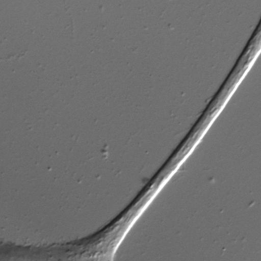
Fig 4. Vesicle Motion in PC12. Movie in QuickTime (7.45MB) and MPEG-1 (1.75MB) format